What is an Eye Exam?
An eye exam is also known as a dilated eye exam or complete eye exam. A normal, complete, comprehensive eye exam is a thorough examination of the eye that includes dilating the pupil of the eye. Ophthalmologists examine the eyes using a variety of tests, equipment, and other methods when other conditions are indicated. A complete eye exam will take around one hour or longer. The dilating eye drops can take some time to fully dilate the pupil.
Who needs an eye exam?
Undergoing a comprehensive eye exam on a consistent basis is important for all adults and children to catch any issues or vision concerns as soon as possible. You should also receive regular eye exams, even if you haven't noticed any changes or concerns with your vision. Men and women who are over 18 and below 40 years old should get their eyes examined every other year. Research has shown us that 25% of students have some kind of vision concern. Ophthalmologists suggest that students have their eyes checked every two years, regardless of whether they appear to have wonderful vision.
Age 20 – 60
You should have a routine eye exam every 3 – 5 years. In your 40s, you will start to develop presbyopia (unable to focus up close) and will need to change your glass prescription every 2 – 3 years.
Age 65 and above
Once you are over 65, you should have a routine eye exam every 1 – 2 years. For people aged 65 and over, the incidence of cataracts, glaucoma, and macular degeneration increases. Unfortunately, many of these diseases do not give symptoms until the disease has caused significant damage to the eye.
Eye Exams Reviews
What to Expect During An Eye Exam
There are a few things that you can expect when arriving at our facility to receive an eye exam. You may also need to provide some information to Dr. Croley and his team so that they can better serve you. When you arrive in the office for your first visit, you will be asked to fill out a health history questionnaire and insurance information. You will need to sign a variety of papers or documents required by the government. The health questionnaire that you fill out gives the office staff an excellent starting point to begin the eye exam. Additionally, our team will need information about the following:
- Chief complaint or history of present eye problem
- Past eye and medical history
- Review of systems
- Social and family history
- List of current medications
This information will allow Dr. Croley and his team will better insight into the current condition of your eyes and past treatments.
Examination Portion of the Eye Exam
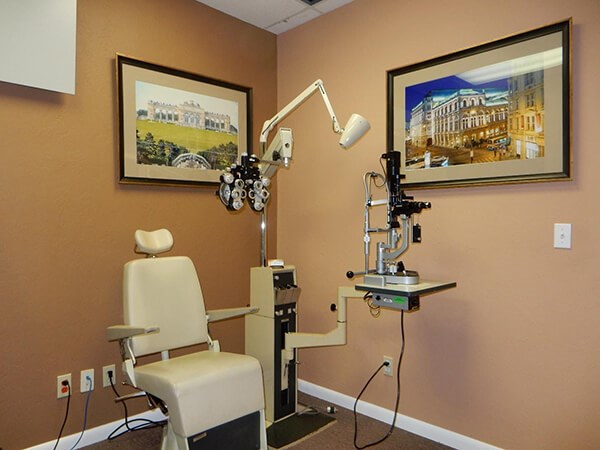 Examination Room
Visual Acuity: The distance vision is tested with a chart that is on the wall. The chart is 20 feet away. Many times, the chart is reflected off a mirror to accomplish the 20 feet so that the exam room doesn’t have to be 20 feet long. A near card may be used to test the near vision. Computers or smart TVs are now used for testing the vision. Color vision may be tested in some circumstances when there is a concern about colored blindness and/or questions about the health of the eye. Pin-hole vision is another way of testing vision. You look through an object with tiny holes in it. Many times, glare testing is used for people that are developing cataracts. This is to determine how much your vision is affected by glare at night. Contrast sensitivity may be used in some cases, which provides more information about the level and quality of a person’s vision.
Eye Movements/Ocular Motility Assessment: The alignment or direction of the eyes is evaluated to see if there is any deviation, such as crossed eyes. The movement of each eye is evaluated by having you follow a light or object and, the quality of how you move your eyes is also evaluated. Your stereopsis or depth perception may be evaluated to see if there are any problems with depth perception.
Pupils: Your pupils are tested to see their reaction to light as this can give an indication of the health of the vision system, including the central nervous system. The pupils also constrict when you look at near objects, which is called accommodation.
Side Vision: The amount of your side vision can be evaluated by a method called confrontation fields. The eye doctor or staff is seated in front of you with you covering one eye. They hold out fingers in the four quadrants of your side vision and ask you to tell them how many fingers do you see. If during the confrontation fields something is found or suspected, a formal visual field examination will be performed with a computerized visual field instrument that is able to find very small changes in your side vision.
External Eye Exam: The ocular adnexa or eyelids and other structures around the eyes are examined for any abnormalities.
Refraction Testing for Eyeglasses: A refraction determines the extent of any myopia (nearsightedness), hyperopia (farsightedness), and/or astigmatism (your eye is shaped like a football cut lengthwise in half instead of a round basketball). Prior to beginning the refraction, you may be taken to a room and your eyes will be tested with a wavefront analyzer (aberrometer) or a computerized autorefractor instrument. These instruments are able to measure your eyes and help determine your eyeglass prescription.
You are brought into a room and placed behind a machine called a phoropter. The phoropter contains all the possible combinations of eyeglass prescriptions. The eye doctor or staff give you choices between two different lenses and asked you which lens do you see better with. The eyeglass prescription is fine-tuned based on your answers to which lens you state that you see the best. This prescription is important as it provides more information than just an eyeglass prescription. Your best correctable vision gives the eye doctor information about the quality of your vision and the health of your eyes. All clinical decisions the eye doctor makes about the health of your eyes is based on your best correctable vision. If your best correctable vision is not 20/20 or it has changed from your prior visit, the eye doctor will need to search for the reason your vision has changed. The eye doctor will order a variety of tests to determine the cause of your vision loss.
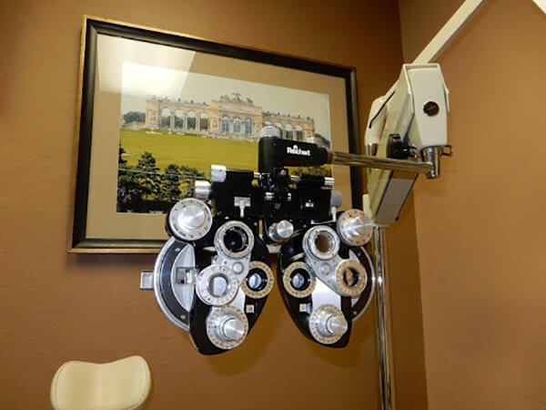 Phoropter
Unfortunately, most insurances do not cover the cost of the refraction. It is a separate fee. It really should be covered as part of an eye exam as it provides important medical information about the health of the eye. But, Medicare and most insurances have taken the easy way out and have chosen not to cover the cost.
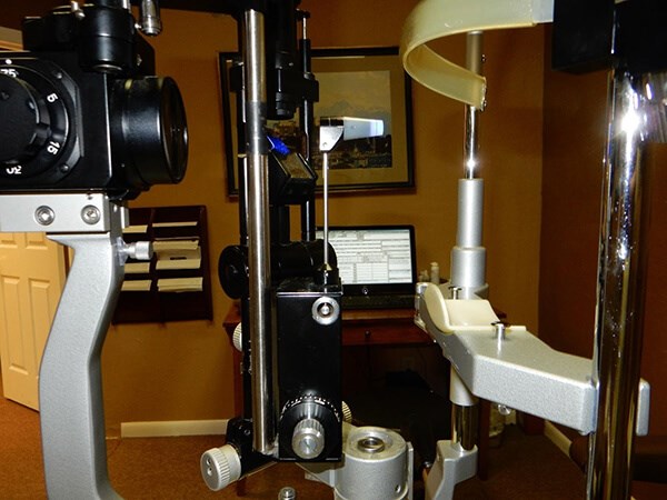
Slit Lamp Exam
Slit Lamp Exam: A slit lamp is a binocular microscope that is used to examine the tissues of the eye under high magnification. You place your chin on a chin rest and place your forehead up against the band attached to the slit lamp. The slit lamp is used to examine the eyelids, conjunctiva, cornea, iris, and lens. Once your eyes are dilated, the slit lamp can also be used to examine the retina and optic nerve inside the eye.
Glaucoma Test, Eye Pressure, or Intraocular Pressure: Your eye pressure will be measured during your eye exam. There are a variety of different instruments that can be used to measure eye pressure:
- Non-Contact Tonometer
A non-contact tonometer means that the instrument does not touch the eye. Another name is the air-puff test, which can measure the eye pressure by using a small burst of air against the cornea. The instrument is able to measure the resistance of the cornea to air applied against the eye.
- Applanation Tonometer
The Goldmann Applanation Tonometer is the gold standard for accurately measuring the pressure inside the eye. It is attached to the slit lamp microscope. An anesthetic or numbing eye drop is placed on the eye, which contains a dye called fluorescein that allows the eye doctor to measure the eye pressure. The tonometer gently presses against the cornea to measure the tension inside the eye. The test is entirely painless as the eye is numb.
Other instruments that can measure the eye pressure are the Tonopen, Icare tonometer, Diaton Transpalpebral tonometer, Pascal tonometer, and Perkins tonometer. It is very important that the eye pressure is measured during the eye exam. Except in cases of a sudden increase in eye pressure as in acute angle glaucoma, you cannot feel that your eye pressure is elevated. Glaucoma does not give any symptoms until a significant loss of side vision has occurred.
Goldmann Applanation Tonometer
Examination of the Retina and Optic Nerve: In order for the eye doctor to be able to see the entire retina, the pupil of the eye needs to be dilated. The pupils are dilated with eye drops that are instilled in the eye. It takes about 20 – 30 minutes or more for the drops to dilate your eyes. The length of time depends on the types of eye drops that are used to dilate the pupils, and dark brown eyes take longer than blue eyes. Once your pupils are dilated, the eye doctor will use a variety of different instruments and lenses to look inside the eye to examine the retina and optic nerve for any sign of eye disease.


Experience Improved Eye Health
After the examination, Dr. Croley will discuss with you the results of the eye exam and provide their recommendations about any diseases or abnormalities found during the exam. It is our goal to help you experience improved eye health and vision clarity. Learn more about scheduling an eye exam by contacting the Cataract & Refractive Institute of Florida today.


